HybriScan Technologies offers the HybriScan Molecular Microscope “HSCMM” consisting of a reflective optical microscope and a state-of-the-art Raman microspectrometer together with an electron microscope. This novel hybrid technology offers nanoscale spatial resolution, information on morphology of materials and exploits the Raman-effect of materials to investigate their chemical and physical properties. The Raman-effect is a non-contact, nondestructive spectroscopic method that uses light to identify and characterize materials on a molecular level.
Relitech collaborated with Hybriscan to extend the Matlab program that is used for their Raman spectroscopy products. The focus on this work was to control a miniature 2D and 3D stage and the coupling of coordinate domains from different image sources, such as from electron microscopy and video microscopy. The GUI of the PC software enables the user to mark certain regions. Below an example of a free form is shown. After calibration, the system scans the selected area by means of a miniature sub-stage.
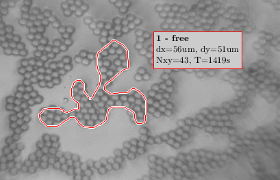
Video image with region of interest (ROI) selected with the interactive GUI.
The substage has been integrated with mounts for easy insertion onto the standard electron microscope stage and also holds Hybriscan’s sample mount. This secondary stage, see below, enables control of the Raman spectrometer with sub-micron precision.
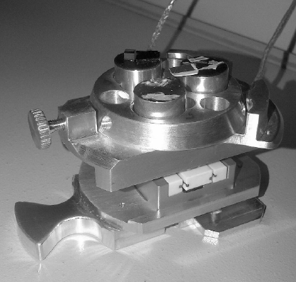
2D substage (travel=20mm) integrated with mount for insertion into the SEM (bottom) and sample mount (top).
Coordinates in sub-micron range with nano meter resolution from a video or scanning electron microscope image are transformed to real world stage coordinates. This makes it possible to acquire spatial Raman spectrum information and to add it to the original image as overlay.
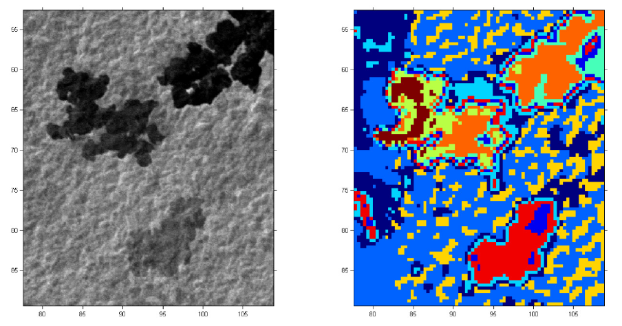
Left a SEM (Scanning Electron microscope) image of a graphene sample. Right the same sample augmented with Raman spectral imaging information.
Besides the regular software development using Matlab, more specialized and non-common aspects of software development can be found within the project:
- The integration of DOT.NET technology within Matlab for the video camera.
- 2D image processing, correlation and affine transformations.
- A third (z axis) linear motor combined with auto focus algorithms.